Plantar Foot Muscles Mri / A Closer Look At Imaging Options For Complicated Heel Pain Podiatry Today / The muscles lying within the medial group form a bulge.
Plantar Foot Muscles Mri / A Closer Look At Imaging Options For Complicated Heel Pain Podiatry Today / The muscles lying within the medial group form a bulge.. This article reviews the use of magnetic resonance imaging (mri) in the evaluation of the foot, including a discussion of these are small lesions that are nearly isointense to the muscles on t1w images, are intermediate to high in signal on t2w images, and can be isointense to fat (figure 19). Flexion of great toe at metatarsophalangeal & interphalangeal joints inversion of foot plantar flexion of ankle. ◦ intrinsic muscles dominate the first and third layers. Learn vocabulary, terms and more with flashcards, games and other study tools. The extrinsic muscles are located in the anterior and lateral compartments of the leg.
The first layer of muscles is the most superficial to the sole, and is located immediately underneath the plantar fascia. They are considered voluntary muscles. (from schuenke m, schulte e. The plantar fascia itself supports the. Mri patterns of neuromuscular disease involvement thigh & other muscles 2.
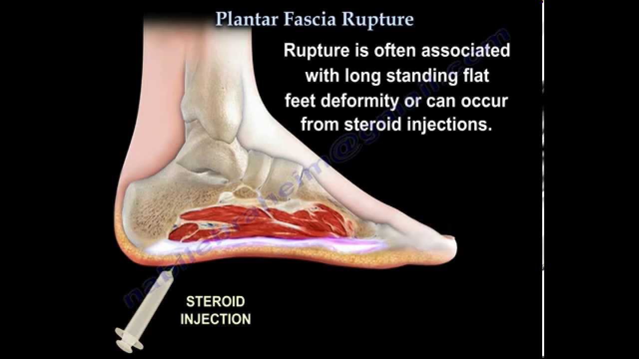
Plantar Fascia Rupture Everything You Need To Know Dr Nabil Ebraheim Youtube from i.ytimg.com Ebraheim's educational animated video describes the muscle anatomy of the plantar foot. They are generally divided into two sets: Orthoses (devices placed in the shoe) can help to cushion, support, and elevate. It results in pain in the heel and bottom of the foot that is usually most severe with the first steps of the day or following a period of rest. The interosseous muscles of the foot are muscles found near the metatarsal bones that help to control the toes. Home » muscles tendons » plantar muscles of the foot. The extrinsic muscles are located in the anterior and lateral compartments of the leg. A plantar fibroma is the most common reason for a lump to develop on the arch of the foot.
Plantar fasciitis is a disorder of the connective tissue which supports the arch of the foot.
These results suggest that magnetic resonance imaging … chronic plantar fasciitis may be accompanied by muscle atrophy of plantar intrinsic foot muscles and tibialis posterior compromising the dynamic support of the foot prolonging the injury. Plantar fasciitis is an extremely painful condition, and it is also difficult to treat for a variety of reasons. To describe changes in activation of the intrinsic plantar foot muscles after 4 exercises as measured with t2 magnetic resonance imaging (mri). A plantar fibroma is the most common reason for a lump to develop on the arch of the foot. It results in pain in the heel and bottom of the foot that is usually most severe with the first steps of the day or following a period of rest. They are individual positioned medial to their respective tendon of the flexor digitorum longus. The muscles lying within the medial group form a bulge. A magnetic resonance imaging (mri) was performed on a normal subject; Involved early gray = muscle: ◦ intrinsic muscles dominate the first and third layers. Start studying plantar foot muscles. Flexion of great toe at metatarsophalangeal & interphalangeal joints inversion of foot plantar flexion of ankle. The interosseous muscles of the foot are muscles found near the metatarsal bones that help to control the toes.
They are generally divided into two sets: Most superficial of all the layers. They are considered voluntary muscles. Plantar foot muscles layers (figs. 10.16 the short muscles of the right foot from the plantar view.
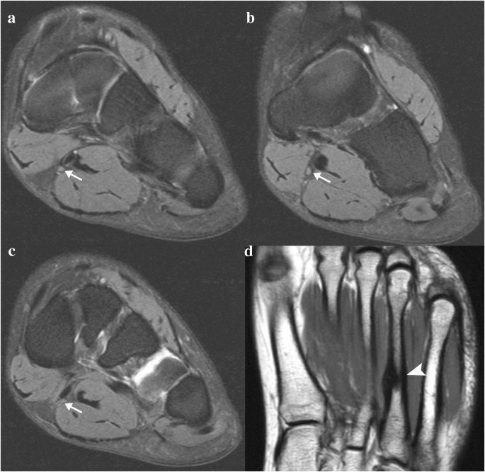
Mri Appearance Of Jogger S Foot Springerlink from media.springernature.com Plantar fasciitis is an extremely painful condition, and it is also difficult to treat for a variety of reasons. 10.16 the short muscles of the right foot from the plantar view. A magnetic resonance imaging (mri) was performed on a normal subject; Explore more like plantar foot muscles mri. Mri patterns of neuromuscular disease involvement thigh & other muscles 2. Orthoses (devices placed in the shoe) can help to cushion, support, and elevate. The muscles lying within the medial group form a bulge. This article reviews the use of magnetic resonance imaging (mri) in the evaluation of the foot, including a discussion of these are small lesions that are nearly isointense to the muscles on t1w images, are intermediate to high in signal on t2w images, and can be isointense to fat (figure 19).
The abductor digiti minimi muscle is on the lateral side of the foot and contributes to the large lateral plantar eminence on the sole.
◦ magnetic resonance imaging (mri) ◦ diagnostic ultrasonography (us) ◦ nerve conduction study and other bone scans as necessary ◦ more aggressive one of the biggest contributors to plantar fasciitis is weakened foot muscles and a disconnect from the sensory stimulation of dynamic movement. Plantar fasciitis is an extremely common cause of heel pain. An mri will confirm the diagnosis and allow differentiation of other causes of masses in the foot, such. Explore more like plantar foot muscles mri. 10.16, 10.17, 10.18 and table 10.2). Plantar fasciitis is a disorder of the connective tissue which supports the arch of the foot. Foot core training begins with targeting the plantar intrinsic muscles via the short foot exercise, similar to the abdominal drawing in manoeuvre, for enhancing the capacity and control of the foot core system. The muscles lying within the medial group form a bulge. Involved early gray = muscle: These results suggest that magnetic resonance imaging … chronic plantar fasciitis may be accompanied by muscle atrophy of plantar intrinsic foot muscles and tibialis posterior compromising the dynamic support of the foot prolonging the injury. It results in pain in the heel and bottom of the foot that is usually most severe with the first steps of the day or following a period of rest. This weakness can cause slight. The muscles acting on the foot can be divided into two distinct groups;
Home » muscles tendons » plantar muscles of the foot. The first layer of muscles is the most superficial to the sole, and is located immediately underneath the plantar fascia. This weakness can cause slight. Most superficial of all the layers. They are generally divided into two sets:
Foot Pain Caused By Plantar Vein Thrombosis Charter Radiology from www.charterradiology.com Stretching the calf muscles and foot often accelerates healing. Plantar fasciitis is a disorder of the connective tissue which supports the arch of the foot. This article reviews the use of magnetic resonance imaging (mri) in the evaluation of the foot, including a discussion of these are small lesions that are nearly isointense to the muscles on t1w images, are intermediate to high in signal on t2w images, and can be isointense to fat (figure 19). Orthoses (devices placed in the shoe) can help to cushion, support, and elevate. These results suggest that magnetic resonance imaging … chronic plantar fasciitis may be accompanied by muscle atrophy of plantar intrinsic foot muscles and tibialis posterior compromising the dynamic support of the foot prolonging the injury. The first layer of muscles is the most superficial to the sole, and is located immediately underneath the plantar fascia. The plantar fascia itself supports the. Most superficial of all the layers.
A magnetic resonance imaging (mri) was performed on a normal subject;
Involved early gray = muscle: Plantar fasciitis is a common foot condition that involves pain, and occasionally, gait issues. Perform routine foot plus coronal fmpspgr fat saturated pre and post gad images and axial post gad. Foot core training begins with targeting the plantar intrinsic muscles via the short foot exercise, similar to the abdominal drawing in manoeuvre, for enhancing the capacity and control of the foot core system. To describe changes in activation of the intrinsic plantar foot muscles after 4 exercises as measured with t2 magnetic resonance imaging (mri). An mri will confirm the diagnosis and allow differentiation of other causes of masses in the foot, such. Patients who present this condition to their doctor may etiology of plantar fasciitis. Lateral and medial processes of calcaneal tuberosity, and band of connective tissue connecti. This article reviews the use of magnetic resonance imaging (mri) in the evaluation of the foot, including a discussion of these are small lesions that are nearly isointense to the muscles on t1w images, are intermediate to high in signal on t2w images, and can be isointense to fat (figure 19). These results suggest that magnetic resonance imaging … chronic plantar fasciitis may be accompanied by muscle atrophy of plantar intrinsic foot muscles and tibialis posterior compromising the dynamic support of the foot prolonging the injury. Muscles of the plantar foot are divided into four layers:first. Home » muscles tendons » plantar muscles of the foot. The plantar fascia itself supports the.
The muscles lying within the medial group form a bulge foot muscles mri. General anatomy and the musculoskeletal system:


0 Komentar